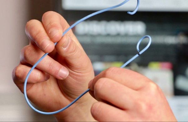
Ureteral Stents: Why Urologists Use Them

Ureteral stents keep the ureters, or tubes that carry urine from the kidneys to the bladder, open. Urinary blockages caused by kidney stones, ureteral stones, constricted ureters, or tumours may necessitate their use. Most ureteral stents are temporary, but some persons with chronic issues require longer-term ureteral stents.
Stents for the ureters are thin, flexible tubes that keep the ureters open. The urinary system includes the ureters. These long, thin tubes usually transport urine from the kidneys to the bladder. Ureteral stents are medical devices that are used to prevent or treat ureteral blockages.
Ureteral stents are 10 to 15 inches long and 14 inches in diameter and are made of silicone or polyurethane (plastic). They line the ureter's whole length, keeping it open. A coil on the top of the stent is placed within a kidney. The lower loop is inserted into the bladder.
What are the benefits of ureteral stents?
Urine can become trapped in the ureters, preventing it from draining normally. A ureteral stent can clear the ureter and allow your kidneys to function normally.Urinary stents are most commonly used to allow urine to flow through the ureter around a kidney stone that is obstructing urine flow. In addition, after breaking up kidney stones, your doctor may use a stent to prevent blockage from the passing fragments. Stents can also be used after kidney stone removal to prevent postoperative edoema from blocking the ureter.
During ureteral stenting, what happens?
Stenting is usually performed as an outpatient surgery. The next day, you return home. The treatment is performed by a urologist, a specialist who specialises in problems that impact the urinary system.You will be given anaesthetic before to the surgery. Most of the time, you'll be sleeping because to general anaesthetic. The treatment is performed while you are lying on your back. Your service provider is:
- To find the obstruction and guide the treatment, doctors use X-ray imaging (fluoroscopy) or a kidney ultrasound.
- Through the urethra and into the bladder, a tiny scope device with a lens (cystoscope) is inserted. When you pee, urine exits your body through the urethra tube.


Comments
Post a Comment